Hydronephrosis
Hydronephrosis
Hydronephrosis means water on the kidneys.
This means that one or both of the baby’s kidneys are stretched and swollen because they are holding on to urine (wee).
Antenatal hydronephrosis is quite common, affecting about 1 in 100 pregnancies. Most cases are not serious. The problem often disappears by the time the baby is born, with no long-term effects on the baby or mother.
You may need more scans during the pregnancy to find out whether the antenatal hydronephrosis continues or causes problems. Your baby may need tests after birth.
Sometimes, antenatal hydronephrosis is caused by other problems, such as when urine refluxes (passes back) towards the kidney, or an anomaly that blocks the flow of urine. Rarely, it suggests more serious problems during the pregnancy or after birth. A few children will need monitoring and/or treatment, such as surgery.

About the urinary system
The urinary system starts to develop several weeks into pregnancy. It gets rid of things that the body no longer needs, so that we can grow and stay healthy.
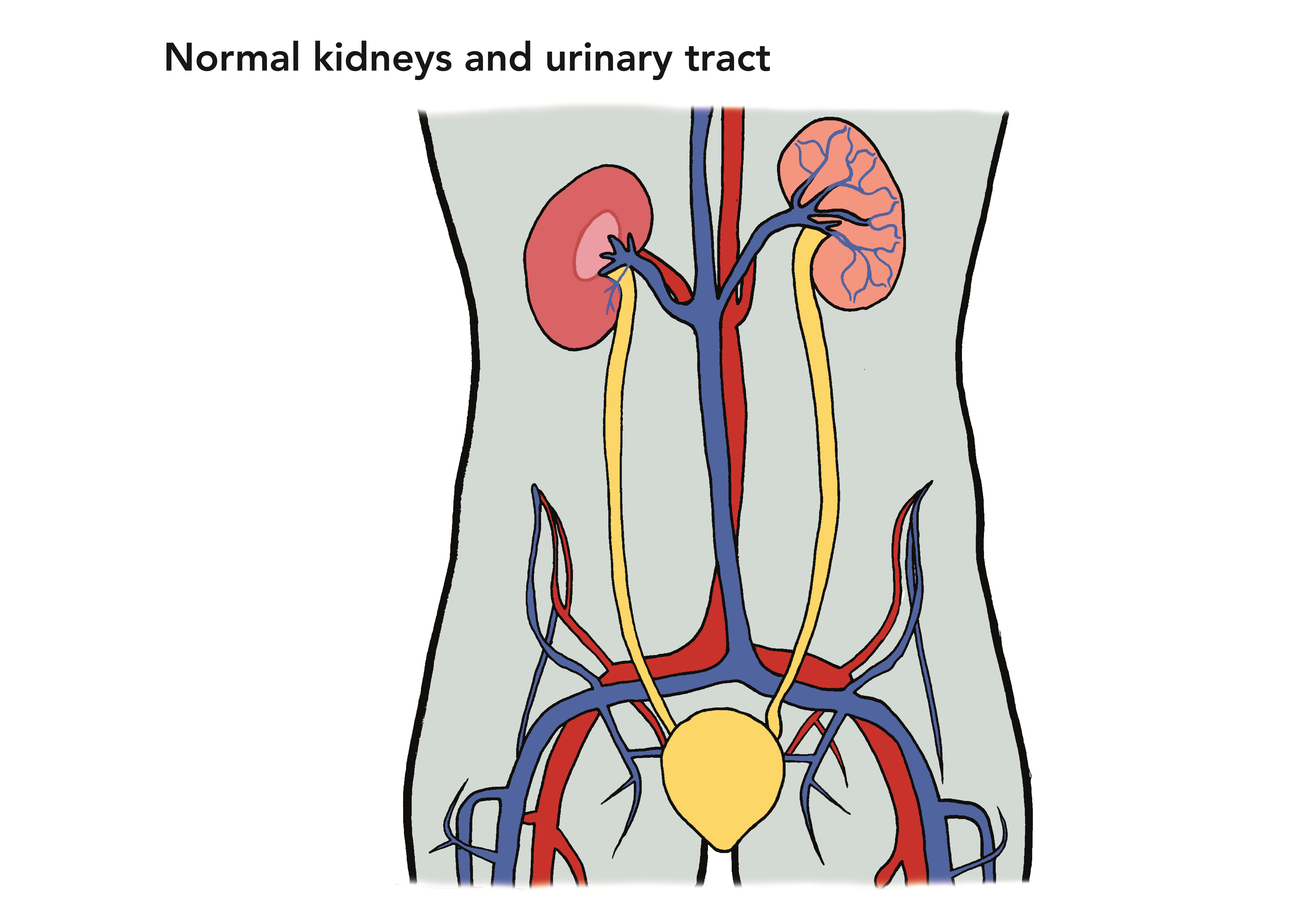
The kidneys are bean-shaped organs. They filter blood to remove extra water and waste in urine (wee). Most of us have two kidneys. They are on either side of our spine (backbone), near the bottom edge of our ribs at the back.
The two ureters are long tubes that carry urine from the kidneys to the bladder.
The bladder is a bag that stores urine until we are ready to urinate (wee). It sits low down in the tummy area.
The urethra is a tube that carries urine from the bladder to the outside of the body.
Causes
Why it happens
Antenatal hydronephrosis is not inherited from the mother or father, and is not caused by anything that the mother does during her pregnancy.
Most cases are not caused by any problems, and get better.
Occasionally, hydronephrosis is caused by other problems:
- when babies with VUR pass urine, some urine refluxes (passes back up) the wrong way towards the kidneys
- an obstruction or ‘blockage’ may partially or fully stop the flow of urine.
- Which parts of the urinary tract are affected?
- The kidney has several distinct parts. After the kidney makes urine, it collects in the renal pelvis before passing into the ureter.
- In antenatal hydronephrosis, the renal pelvis holds on to urine and it swells up. Sometimes, the ureter that is connected to the kidney also swells up.
- No other problems
- In most cases, there are no other problems with the urinary system, and the antenatal hydronephrosis usually resolves (gets better) before birth. These children will probably not have any long-term problems and will not need follow-up after birth. Doctors think antenatal hydronephrosis with no other problems happens as the baby’s urinary system develops – especially if one or both ureters do not fully develop before the kidneys start making urine.
- Reflux
- Some cases of antenatal hydronephrosis are caused by vesicoureteral reflux (VUR). In VUR, some urine refluxes (goes back up) towards one or both kidney. In severe VUR, the reflux reaches into the kidney, causing the antenatal hydronephrosis.
- Blockage
- Some cases are caused by a blockage (or obstruction) that partially or fully stops the flow of urine out of the kidney. If this is suspected, you may be referred to a paediatric urologist (a surgeon who treats children with problems of the urinary system), who will monitor your child.
- There are different types of problems that cause a blockage. These may be diagnosed after a baby is born, and may need treatment.
- Posterior urethral valves (PUV): some boys are born with extra flaps of tissue in part of the urethra, the tube that carries urine out of the body. A long, thin tube (catheter) is inserted into the urethra to drain urine. PUV are removed by surgery.
- Pelviureteric junction (PUJ) dysfunction (or PUJ obstruction): a blockage between the renal pelvis (in the kidney) and the ureter, the tube that carries urine from the kidney to the bladder. In some cases, surgery is needed.
- Vesicoureteric junction (VUJ) dysfunction (or VUJ obstruction): a blockage between the ureter and the bladder. Many cases get better on their own, but in some cases, surgery is needed.
- Duplex kidney: sometimes a kidney and ureter on one side of the body develop in two parts. In some cases, one of the ureters dilates (swells) where it goes into the bladder – this is called a ureterocele. In many cases, this will not need follow-up or treatment, but if this causes blockage or reflux, children may need more tests and occasionally surgery.
Tests and Diagnosis
Before birth
In antenatal hydronephrosis, the part of the kidney that swells is the renal pelvis. The 20 week antenatal ultrasound scan looks at the baby growing in the womb and measures the size of the renal pelvis.
What the ultrasound can tell us
The ultrasound cannot give precise information about the problem or what is causing it. Although your doctor will not always know how your baby will be affected at birth, he or she is less likely to have serious problems if:
- he or she is growing well in the womb
- no other problems have been found
- the amount of amniotic fluid (or liquor), which is the liquid that the baby is floating in, is normal.
Often, the expectant mother will need more ultrasound scans during her pregnancy. These will check whether the problem goes away or gets worse.
Referral
Your obstetrician may refer you to specialist healthcare professionals, such as a paediatric nephrologist (a doctor who treats children with kidney problems) or a paediatric urologist (a surgeon who treats children with problems of the urinary system).
You can speak with these doctors about possible complications and their management.
After birth
You may need to come back to the hospital for your baby to have imaging tests (scans). These use special scanners that take pictures of the inside of his or her body.
Ultrasound scan
The first test is normally an ultrasound scan, which is similar to the scan mothers have in pregnancy. It looks at the shape and size of the kidneys and other parts of the urinary system. A small handheld device is moved around your baby’s skin. A machine attached to the probe directs sound waves into his or her body – your child cannot feel these. These sound waves are turned into pictures that can be seen on a screen.
Other tests
- Your doctor may also arrange a DMSA scan, which checks for any damage in the kidneys, and/or a MAG3 scan, which shows whether blood is going into the kidneys and whether there is a blockage in the urinary system. These are normally done when a baby is some weeks old. In each test, a chemical that gives out a small amount of radiation (energy) is injected into one of your baby’s blood vessels – a special gel or cream can be used to stop your baby feeling any pain. A special camera takes images (pictures) of your baby’s kidneys.
- An MCUG (sometimes called a VCUG) checks how your baby is passing urine, and whether there is any reflux (when urine passes back up towards the kidneys). A thin flexible tube called a catheter is passed through your baby’s urethra and a dye is put through to reach the bladder – this does not hurt your baby. A special X-ray machine takes a series of images of your baby’s bladder while it is emptying.
Treatment
Before birth
Treatment before birth is very rare. It is normally only recommended when there is little or no amniotic fluid (or liquor), the liquid that surrounds the baby growing in the womb. Your healthcare team will speak with you about this.
After birth
Many babies do not need treatment after birth. This depends on findings from the antenatal ultrasound scans and tests after birth. In most cases, babies can be discharged home a short time after birth.
Rarely, babies are born with complications and need to be moved to a neonatal unit, an area of the hospital for newborn babies, for monitoring and treatment.
Preventing and treating UTIs
Some babies are at higher risk of urinary tract infections (UTIs). If they keep coming back, they may cause damage to a kidney. To protect your child’s kidneys, it is important to prevent UTIs, and treat them quickly when they do happen.
- Your baby may need to take a small dose of antibiotic medicine each day – this is called a prophylactic antibiotic. Antibiotics kill the bacteria (germs) that cause UTIs, and so help prevent these infections.
- Sometimes UTIs can happen even when your child is taking these antibiotics. It is important that UTIs are diagnosed and treated quickly to try to prevent them causing kidney damage. If you think your child has a UTI, contact your doctor or local NHS services.
It is important to follow your doctor’s instructions about when and how much to give. Continue to give the antibiotics to your baby as your doctor has told you.
Surgery
A small number of babies who had antenatal hydronephrosis in the womb have complications after birth. These babies may be monitored or treated in a neonatal unit, a special area of the hospital for newborn babies.
Occasionally, a baby needs an operation to correct the problem that is causing the hydronephrosis.
